


Understanding where a drug distributes in vivo remains a critical, yet often underestimated, challenge in preclinical development. Many programs still rely on blood-based PK or ex vivo tissue analysis, generating incomplete or fragmented views of biodistribution. These approaches increase animal use, limit longitudinal insight, and can introduce risk by leaving key questions around target engagement and off-target exposure unanswered.
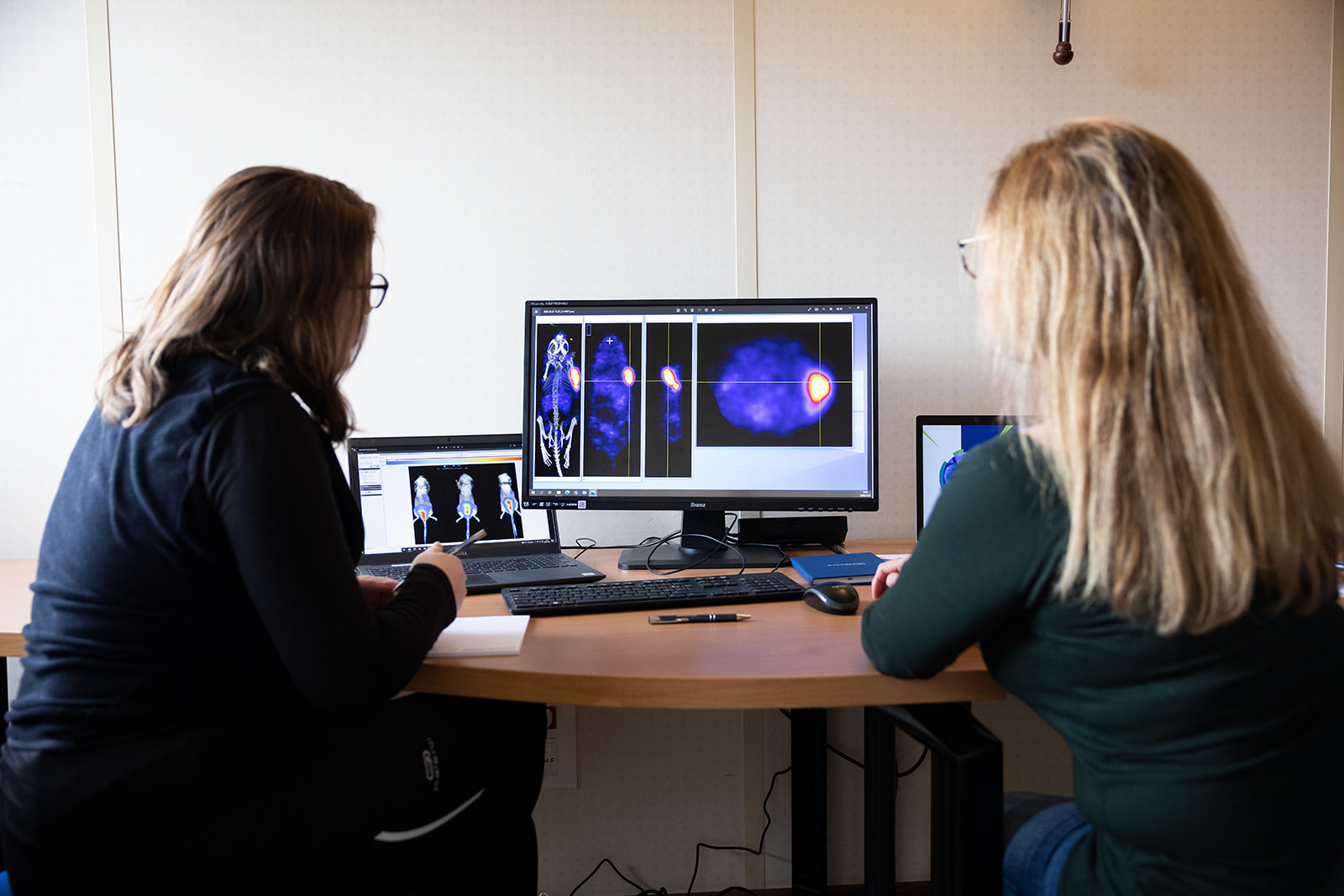
In this webinar, we will explore how in vivo imaging can provide a more complete and efficient view of biodistribution. You will gain a practical understanding of when to apply optical imaging approaches such as fluorescence and bioluminescence, including their strengths for rapid screening and multi-timepoint assessment, while supporting the reduction of research models through longitudinal study design.
We will also examine quantitative nuclear imaging techniques, including PET and SPECT, and how they compare to optical techniques in terms of sensitivity, precision, and study design considerations. Through this comparison, participants will learn how to select the most appropriate approach based on their specific program needs, while aligning biodistribution strategies with evolving 3Rs expectations and regulatory focus.
Key Learning Objectives
- Understand the limitations of conventional biodistribution approaches and their impact on translatability.
- Identify when to apply optical versus nuclear imaging techniques based on study objectives and constraints.
- Review how imaging can support longitudinal study design, reducing and refining the requirement for animal models in biodistribution studies.
Join this webinar to explore how imaging can generate more informed biodistribution data and better align preclinical programs with 3Rs principles.
